Defining the neuroimmune-cancer cell axis
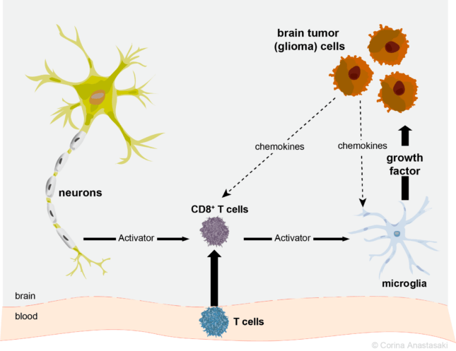
For the past 25 years, my laboratory has developed and employed genetically engineered mice to understand why brain tumors form in children with the Neurofibromatosis type 1 (NF1) cancer predisposition syndrome1. Using these authenticated preclinical mouse models of NF1-associated optic gliomas (low-grade astrocytomas of the optic nerves)2, we showed that microglia, macrophage-like cells in the brain, produce growth factors necessary for tumor development, proliferation, and vision loss3, 4, 5, 6. However, it was not clear how microglia get activated to control tumor pathogenesis.
This all changed thanks to Reviewer #3.
To create a transplantable low-grade glioma model, Dr. Yi-Hsien Chen, a postdoctoral fellow in our group, isolated cancer stem cells from Nf1 optic glioma mice, and showed that they could form tumors when injected into the brains of naïve immunocompetent mice7. We were very excited about this finding, and submitted the paper for review. We were surprised that one of the reviewers insisted that we repeat these experiments using immunocompromised mice, similar to what other investigators have historically employed for human brain tumor xenograft studies. At first blush, this seemed like a strange request, as we had already demonstrated tumor formation in mice with a normal immune system. To our amazement, when Yi-Hsien injected these optic glioma stem cells into athymic mice lacking functional T cells, no tumors were observed!
The finding that T cells were required for brain tumor formation was exciting, and prompted another postdoctoral fellow, Dr. Yuan Pan, to confirm and extend these observations. Yuan went on to demonstrate that the inability of athymic mice to support optic glioma formation resulted from impaired microglia function, including reduced expression of Ccr2 and Ccl5, both of which are required for Nf1 optic glioma growth. She further demonstrated that reduced Ccr2 and Ccl5 expression by athymic microglia could be restored by soluble factors produced by activated T cells8.
While these studies established a critical role for T cells in microglia-mediated glioma formation and growth, how T cells became activated and how they created a supportive tumor microenvironment remained unknown. Enter Dr. Xiaofan (Gary) Guo, a graduate student in our laboratory who had previously shown that T cells and microglia are drawn into the tumor microenvironment by chemokines secreted from the glioma cells themselves9. In our latest report10, Gary and his colleagues nicely demonstrated that T cell entry into the brain is required for optic glioma growth, and that, upon activation, T cells produce Ccl4, which in turn induces microglia to secrete Ccl5, the growth factor necessary for optic glioma growth11. Moreover, Gary discovered that neurons were the cells responsible for activating T cells, thus establishing a neuroimmune-cancer cell circuit.
We are very excited about these findings, as they have important implications for the field.
First, the fact that nerve cells are active participants in brain tumor development and growth adds to our growing appreciation of the role of neurons in cancer – ushering in the new field of Cancer Neuroscience12. Researchers in our team are now investigating the role of neurons in dictating human and mouse nervous system tumor formation and progression. Second, our studies demonstrate that T cells are key regulators of the brain tumor microenvironment, prompting us to explore the idea that T cells can alter microglia function in the brain, both in health and in the setting of central nervous system disease13. Third, since we showed that T cells are recruited into the optic nerve (brain) from the blood, we hypothesize that T cells may serve as integrators of risk factors for brain disorders, including gliomas. Current studies in the laboratory are focused on understanding how environmental exposures and systemic diseases impact on brain tumor formation and progression in mice14, 15.
I want to thank the wonderful trainees whom I have had the pleasure of mentoring, and look forward to similarly exciting new breakthroughs from future members of our team. You can learn more by following us on Twitter (@GutmannLab) and the Gutmann laboratory website (https://gutmannlab.wustl.edu).
Learn more about Tumor Microenvironment & Immunology
Former Laboratory Tumor Microenvironment Researchers
Yi-Hsien Chen, PhD 
Yuan Pan, PhD 
XIaofan Guo, MD, PhD
Current Laboratory Tumor Microenvironment Researchers

Corina Anastasaki, PhD 
Jit Chatterjee, PhD 
Amanda de Andrade Costa, MS 
Olivia Cobb, MS
References
- Campen CJ, Gutmann DH. Optic Pathway Gliomas in Neurofibromatosis Type 1. J Child Neurol 33, 73-81 (2018).
- Hegedus B, et al. Preclinical cancer therapy in a mouse model of neurofibromatosis-1 optic glioma. Cancer Res 68, 1520-1528 (2008).
- Daginakatte GC, Gianino SM, Zhao NW, Parsadanian AS, Gutmann DH. Increased c-Jun-NH2-kinase signaling in neurofibromatosis-1 heterozygous microglia drives microglia activation and promotes optic glioma proliferation. Cancer Res 68, 10358-10366 (2008).
- Daginakatte GC, Gutmann DH. Neurofibromatosis-1 (Nf1) heterozygous brain microglia elaborate paracrine factors that promote Nf1-deficient astrocyte and glioma growth. Hum Mol Genet 16, 1098-1112 (2007).
- Pong WW, Higer SB, Gianino SM, Emnett RJ, Gutmann DH. Reduced microglial CX3CR1 expression delays neurofibromatosis-1 glioma formation. Ann Neurol 73, 303-308 (2013).
- Toonen JA, Solga AC, Ma Y, Gutmann DH. Estrogen activation of microglia underlies the sexually dimorphic differences in Nf1 optic glioma-induced retinal pathology. J Exp Med 214, 17-25 (2017).
- Chen YH, et al. Mouse low-grade gliomas contain cancer stem cells with unique molecular and functional properties. Cell Rep 10, 1899-1912 (2015).
- Pan Y, et al. Athymic mice reveal a requirement for T-cell-microglia interactions in establishing a microenvironment supportive of Nf1 low-grade glioma growth. 32, 491-496 (2018).
- Guo X, Pan Y, Gutmann DH. Genetic and genomic alterations differentially dictate low-grade glioma growth through cancer stem cell-specific chemokine recruitment of T cells and microglia. Neuro Oncol, 21, 1250-1262 (2019).
- Guo X PY, Xiong M, Sanapala S, Anastasaki C, Cobb O, Dahiya S, Gutmann DH. Midkine activation of CD8+ cells establishes a neuron-immune-cancer axis responsible for low-grade glioma growth. Nat Commun In press, (2020). https://rdcu.be/b3Ud
- Solga AC, et al. RNA Sequencing of Tumor-Associated Microglia Reveals Ccl5 as a Stromal Chemokine Critical for Neurofibromatosis-1 Glioma Growth. Neoplasia 17, 776-788 (2015).
- Monje M, et al. Roadmap for the Emerging Field of Cancer Neuroscience. Cell 181, 219-222 (2020).
- Wright-Jin EC, Gutmann DH. Microglia as Dynamic Cellular Mediators of Brain Function. Trends Mol Med 25, 967-979 (2019).
- Johnson KJ, Zoellner NL, Gutmann DH. Peri-gestational risk factors for pediatric brain tumors in Neurofibromatosis Type 1. Cancer Epidemiol 42, 53-59 (2016).
- Porcelli B, Zoellner NL, Abadin SS, Gutmann DH, Johnson KJ. Associations between allergic conditions and pediatric brain tumors in Neurofibromatosis type 1. Fam Cancer 15, 301-308 (2016).
Originally published on May 01, 2021 in Nature Portfolio Cancer Communications